Department of Neurosciences
The department of neurosciences provides specialized care in the diagnosis and treatment of all neurological conditions and includes speciality services from pediatric neurosurgery, pediatric neurology, neuro interventional radiology, neuropathology, neuroradiology, neuro-critical care, cranial and facial surgery, and neuropsychology, in addition to other specialists that may be involved in the management and care of children with complex medical diagnoses.
The department of neurosciences aims to provide comprehensive care and support to all patients and families that are dealing with a neurological disease. Involvement of physiotherapy, occupational therapy, speech and language pathology, dietician, child life specialist, social work, and orthotics are an integral part of our approach to provide the best personalized care for children under our care.
Conditions We Treat
Pediatric Brain Tumors
Pediatric brain tumors are the most common type of solid tumors that occur in children. Children with these diagnoses are treated by a specialized team at El Paso Children’s Hospital including a pediatric neurosurgeon, pediatric intensivist, medical oncologist, and radiation oncologist. Management may include close observation, biopsy, or surgical removal based on multiple factors such as appearance on imaging, likely diagnosis, and clinical presentation. Malignant pediatric brain tumors contain cancer cells and can be progressive causing symptoms such as headaches, vomiting, walking difficulty, large heads, and problems with vision or the nerves that control eye movement. These types of tumors require urgent diagnosis and treatment to achieve the best outcomes and may require further surgeries, chemotherapy or radiation therapy based on the type of tumor and age of the child. Benign or noncancerous tumors have several types as classified by the World Health Organization (WHO).
Pediatric brain tumors are different than those that occur in adults and require specialized pediatric care. Most children diagnosed with a pediatric brain tumor go on to lead healthy lives, however, appropriate diagnosis, treatment and follow up is needed at specialized pediatric cancer centers such as El Paso Children’s Hospital. We assess children in our specialized pediatric neurosurgery and oncology clinics, imaging often includes MRI scans of the brain to identify the exact location, shape, size, and chemistry of the tumor. With brain mapping technology used to identify critical surrounding structures to individualize the surgical approach for each patient to preserve neurological function, 3D navigation technology is used during surgery for submillimeter accuracy, and monitoring of neurological function during surgery provides additional details to provide the highest level of care of all children with brain tumors. After surgery, rehabilitation services are involved if needed to help with ongoing recovery. Tumors undergo molecular, immunohistochemistry, and genetic analysis to identify the type and subtype for targeted therapy. Radiation therapy is sometimes needed for certain types of brain tumors and also depends on the child’s age.
As a member of Children’s Oncology Group, pediatric patients with brain tumors have access to the latest clinical trials and research advances. Our pediatric neurosurgeon, oncologist, pathologist, neurologist, psychologists, psychiatrists, nurses and social workers, work together to achieve the best possible medical outcome and quality of life for each pediatric brain tumor patient.
Pediatric Spine Tumors
Pediatric spine tumors can present with back pain, pain radiating to the arms or legs, sensory changes, difficulty walking, weakness in the legs, and changes in bowel and bladder control. Some pediatric spine tumors can grow over a few months or years and lead to progressive and sometimes permanent neurological problems; therefore, early diagnosis and treatment is needed. Treatment can include surgical decompression to relieve the pressure on the spinal cord and/or nerves, surgical resection of the tumor, and sometimes placement of instrumentation along the spinal column if the tumor causes instability of the spine.
Symptoms & Causes
Chiari 1 malformation (CM1) occurs when the cerebellar tonsils (the lowest part of the cerebellum in the back of the head) sinks lower down into the spinal canal. Children diagnosed with CM1 often have no signs or symptoms. They can be diagnosed incidentally after having a CT or MRI scan of the brain done for another problem. Most cases do not require any surgery and children are followed for any symptoms or signs associated with CM1. In those with symptoms, the flow of cerebrospinal fluid (CSF) which flows from around the brain into the spinal canal is blocked by the CM1 causing symptoms to develop. Symptoms most often start in the teen years or early adulthood and can include: headaches involving the back of the head and neck, especially when coughing, sneezing, or straining, and less commonly balance problems, dizziness, trouble swallowing, numbness or tingling in the arms or legs, vision problems, breathing problems, especially during sleep, loss of bowel or bladder control, and a curve of the spine called scoliosis. It is less common to occur in infants and toddlers but symptoms can include trouble swallowing, irritability, and breathing problems. After assessment by a pediatric neurosurgeon and appropriate imaging, often with an MRI of the brain and spine, children with CM1 that have symptoms affecting their day-to-day quality of life may be candidates for surgery to remove the bone pressing against the cerebellar tonsils (decompression) and enlarging the space around the tonsils (duraplasty) to re-establish CSF flow and relieve the symptoms.
Chiari 2 malformations (CM2) are another more severe congenital malformation. Children with type II Chiari malformations are almost always associated with a spinal defect known as myelomeningocele, and are more likely to have symptoms associated with increased pressure inside the brain and on the brainstem. These may include: a build‐up of fluid in the brain (hydrocephalus), a weak cry, trouble swallowing, breathing problems, problems with nerve function in the throat and tongue. Treatment often includes repair of the spinal defect soon after birth and placement of ventricular peritoneal shunt or endoscopic treatment in select cases to relieve the pressure on the brain if symptoms and signs of hydrocephalus are present.
Craniosynostosis occurs when one or more sutures (a natural opening in the skull bone to allow growthof the skull and brain as the child grows) closes prematurely. This causes compensatory growth along the other sutures and no growth along the fused suture causing an abnormal shape of the head and can be associated with increased pressure inside the head in some children. Some craniosynostosis cases are associated with genetic syndromes and may involve multiple sutures or have other medical conditions (see Cranial and Facial Program for more details).
Hydrocephalus occurs when cerebrospinal fluid (CSF) accumulates in the brain and spinal canal, causing the heads of newborns and young children to enlarge and puts pressure on sensitive brain tissue. Hydrocephalus can be congenital, occur after a bleed or infection in the brain, after a major traumatic brain injury, or due to a cyst or tumor. If not treated, hydrocephalus can lead to serious health and cognitive problems. Symptoms of hydrocephalus vary depending on a child’s age and how advanced the condition is when it is discovered. Infants with hydrocephalus may have: an unusually large head (macrocephaly), a rapid increase in head size, extreme sleepiness, vomiting that is frequent and severe, trouble looking up when the head is facing forward, seizures that have no known cause. Older children with hydrocephalus may have: severe headaches, often with nausea and vomiting, blurred or double vision, problems with balance, trouble looking up when the head is facing forward, problems with coordination, trouble standing or walking, loss of bladder control, extreme tiredness, irritability for no apparent reason, delays in reaching developmental milestones, trouble remembering and focusing, and sudden changes in personality. Treatment for hydrocephalus is individualized for each child based on the cause and anatomy of the brain. Options may include medications for cases such as idiopathic intracranial hypertension, endoscopic fenestration procedures for cysts, endoscopic third ventriculostomy (ETV) and choroid plexus cauterization (CPC), or placement of an internal shunt system for continuous drainage of CSF. Our pediatric neurosurgeon assesses each child’s individual case, reviews the imaging, and discuss the best options with families to achieve the best long-term outcomes.
What is spina bifida?
A congenital disorder that occurs early in pregnancy and can lead to a number of problems for the newborn. They range from being born with an open defect along the back with exposed spinal cord and nerves (myelomeningocele) to a normal looking back with internal problems with the spinal cord and nerves (meningocele or closed neural tube defects). Our multispecialty Spina Bifida clinic includes a pediatric neurosurgeon, pediatric urologist, pediatric orthopedic surgeon, case worker, physiotherapist, orthosis specialist, and clinic coordinator to provide individualized care for each child and their needs. Tethered cord is a condition where the filum terminale (a structure located at the end of the spinal cord to anchor it) is thickened and contains fat cells that prevent the spinal cord from elongating as your child grows causing injury to the nerves. If this occurs, it can cause tethered cord syndrome and can lead to weakness in the legs, sensory loss, changes in bowel or bladder control, recurrent urinary tract infections, tip-toe walking, in-toeing of the feet, scoliosis, or back and leg pain. Management involves appropriate imaging of the spine, assessing the function of the bladder, and once diagnosis is confirmed, surgical cutting of the thickened filum through a small opening in the lower back to relieve the tension on the spinal cord and nerves and prevent progressive injury to the neural tissue.
What to Expect:
Treatment and Recovery
If diagnosed before birth with ultrasound or fetal MRI, our specialized fetal, neonatal, and pediatric neurosurgery team review each case, and meet with the parents for counselling to discuss the diagnosis, options for fetal surgery treatment (surgery while in the womb) and appropriate referrals if meeting criteria, and what to expect during pregnancy and after delivery. At birth, if the defect is open, our pediatric neurosurgeon proceeds to surgically repair and close the exposed nerves within 24 to 48 hours. After the patient is discharged, any problems that develop with the kidneys or bladder are treated by our pediatric urologist while any orthopedic related problems are treated by our pediatric orthopedic surgeon. As part of the multidisciplinary clinic, physical and occupational therapy is provided by experienced therapists. Our social service team provides help to the family to navigate the complex medical process. All of this work is done in a coordinated fashion, in one visit, by our highly qualified medical team.
First Visit
During your child’s first visit to the Spina Bifida Clinic, your child will be evaluated by a team of highly experienced surgeons, therapists and clinical staff. Our team members will provide your child with a personalized evaluation and plan of treatment.
Follow Up
Upon follow‐up you will review imaging studies with your providers. We will evaluate any new orthotics. We will continue with routine follow‐up assessments with neurosurgery, orthopedics, urology, physical therapy and social work. We will discuss further recommendations and help develop treatment plans.
Cysts can occur inside the central nervous system (CNS) and can include arachnoid cysts some of which remain stable and only require routine imaging while others can grow and cause increased intracranial pressure and require treatment through minimally invasive endoscopic approaches or placement of a shunt. Other cysts that can occur in the brain or spine include epidermoid and dermoid cyst that grow overtime or can rupture and cause chemical meningitis and often require surgical resection. Rare cysts such as neuroenteric cysts can cause pressure on surrounding structures and require removal. Other cysts that can occur include post-traumatic, post-hemorrhagic (after bleeding inside the brain or spine) or post-infectious (after CNS infections such as meningitis), and can sometimes require specialized treatment.
Children can sometimes be born with or develop vascular malformations such as Vein of Galen malformations, arterio-venous malformations, cavernous malformations, cerebral aneurysm, capillary telangiectasias, sinus pericrania, and dural arterio-venous fistulas. These can present with cardiac problems, seizures, acute neurological changes, headaches, or discovered during imaging for certain genetic mutations. These require a specialized multi-disciplinary approach by our pediatric neurosurgeon and interventional neuro-radiologist for appropriate workup, counselling, and treatment of these conditions based on each individual case. These can include observation, surgery, endovascular treatment (coils or embolization), radiation, or a combination of these.
Strokes and intracranial bleeds are fortunately rare in children but when they occur require urgent diagnosis and appropriate treatments to prevent further damage to the brain. Strokes can include sinus thrombosis (blood clots in the veins that drain the brain) and are treated with blood thinners, other causes include arterial thrombi or emboli that may require clot busters or endovascular treatment to re- establish blood flow to the brain. Moyamoya disease and syndrome occur when the arteries at the base of the skull have narrowed making the brain more susceptible to strokes and bleeds and is treated with bypass surgery by a neurosurgeon. Intracranial bleeds can occur due to tumors, vascular malformations, medications or trauma and when severe require urgent evacuation of the clot through surgery. Our pediatric neurosurgeon, pediatric neurologist, interventional neuroradiologist, and hematologist will often be involved in the care and management of these patients.
Skull tumors can occur in isolation or as part of spectrum of diseases and range from benign lesions such as dermoid cysts, inflammatory disease such as Langerhans histiocytosis, to bony tumors many of which will require biopsy for diagnosis and if needed surgery to remove the lesion and reconstruct the skull defect with bone or artificial protective material.
Congenital defects of the skull can cause herniation of the dura (meningocele) or brain (encephalocele) and often occur in the midline anywhere from the nasal cavity to the back of the head. These require appropriate imaging to identify the structures within the herniated tissue and are often treated by removal of the herniated tissue that is non-functional, closure of the defect, and reconstruction of the skull deformity. This is done by our pediatric neurosurgeon and occasional in collaboration with our craniofacial, ophthalmology, or ear, nose, and throat surgeons based on the location and size of the defect with the use of 3D reconstructions if needed for better surgical planning.
Skull fractures are frequent and occur after falls or blunt trauma to the head. These often require observation to ensure the bone is healing without any complications. In younger children, monitoring for rare growing skull fractures where the dura (lining of the brain) is caught between the fracture pieces and can increase in size over time, these therefore require surgery to repair the defect.
There are many congenital malformations of the brain and spine that can occur. When diagnosed in utero (during pregnancy) we are able to provide counseling to the parents about expectations and post-delivery care. Those discovered after birth can include developmental anomalies of the brain and spine, some of which can be associated with intellectual challenges, seizures, hydrocephalus, hormone deficiencies, visual problems, all of which require appropriate diagnosis and management. At times, we may request genetic testing if a particular syndrome or chromosomal anomaly is suspected.
Congenital malformations of the spine can include spina bifida, vertebral anomalies, scoliosis or kyphosis, basilar invagination and impression, split cord malformation, and others. These require appropriate imaging and clinical examination to establish the best treatment which may include bracing, traction, or surgical correction and stabilization.
Epilepsy is a common nervous system disorder. It is a neurological condition involving the brain that causes seizures. Pediatric epilepsy affects 1% to 2% of children worldwide. A child is said to have epilepsy if they have two or more seizures on separate days. There are many different causes of epilepsy, many of which can be treated with medication alone. However, 1 in 5 children can develop seizures that are not responsive to medications. Seizures cause ongoing damage to the brain and significantly affects the child’s quality of life during development and into adulthood. Surgery plays an important role in children with medically refractory epilepsy and focal seizures where it may helps decrease seizure severity and frequency and at times even be curative. Children with seizures should be assessed by our pediatric neurologist who will often order an electroencephalogram (EEG) to identify which part of the brain the seizure is coming from and a CT or MRI of the brain to rule out any lesions causing the seizures. If surgery is being considered, then further investigations can include specialized CT scans, functional MRIs, cognitive tests, and more invasive monitoring including stereotactic electroencephalogram (SEEG) or strip electrodes to identify with better precision the area and extent of seizure activity. Surgical treatment for epilepsy includes resection of the epilepsy focus, corpus callosotomy, Vagus nerve stimulators (VNS), deep brain stimulation (DBS), and responsive neurostimulators (RNS) and are chosen after discussion with the patient and their family based on the cause and location of the seizures, investigation results, and is unique to each individual child and their seizure to achieve the best outcomes, as one child’s treatment may not be the right treatment for another patient.
Trauma to the brain and spine requires management at specialized centers such as El Paso Children’s Hospital with a trauma team that includes trained trauma specialists, neurosurgeons, and emergency care physicians all certified in the management of pediatric advanced life support to rapidly stabilize and appropriate managed pediatric trauma and provide the best possible outcomes for these patients. Our physiotherapist and occupational therapists are involved once the acute phase is over and recovery has begun to work with each patient to provide the appropriate assessment, tools, and interventions needed to optimize recovery. Patients that may benefit from further neurological rehabilitation are sent to specialized pediatric rehab centers.
Our Brachial Plexus speciality clinic includes assessment by our plastic surgeon and pediatric neurosurgeon for birth related injuries to the peripheral nerves, e.g. brachial plexus injuries, and also treatment of peripheral nerve injuries due to trauma or tumors.
Children with cerebral palsy and other neurological disorders may suffer from spasticity of the limbs or dystonia. This can affect the function and quality of life of the child. At El Paso Children’s Hospital, we offer selective dorsal rhizotomy (SDR), a procedure that targets abnormal sensory nerve roots in the spine and through disconnection of this circuit relieves the spasticity in the legs. Other options for treatment of spasticity include medications, Botox injections into select muscles with spasticity, and baclofen pumps. We work closely with our orthopedic surgeons and orthosis specialists for children with hip dislocations, joint contractures, scoliosis, and short ligaments and tendons for appropriate surgeries to help improve functionality.
Cranial and Facial Program
The Cranial and Facial Clinic at El Paso Children’s Hospital is the first and only comprehensive pediatric cranial and facial program in the region that provides children in the greater El Paso area access to a dedicated multidisciplinary team of physicians skilled in correcting facial, jaw and skull abnormalities. It is the only Cranial and Facial Team in the region to be nationally recognized by the American Cleft Palate‐Craniofacial Association. It is no longer necessary for families to bear the burden of traveling outside of the region to access this service, and our team provides family‐centered, compassionate care you’ve come to expect from a dedicated children’s hospital.
Conditions we treat:
- Craniosynostosis (isolated and syndromic)
- Rare craniofacial clefts
- Acute and chronic sequelae (condition resulting from a disease, injury or other trauma) of facial fractures
- Micrognathia (undersized jaw)
- Apert Syndrome (a kind of dentofacial deformity)
- Crouzon Syndrome (another kind of dentofacial deformity)
- Pfeiffer Syndrome (another kind of dentofacial deformity)
- Treacher Collins Syndrome
- Hemifacial Microsomia (small or underdeveloped parts of the face)
- Parry‐Romberg Syndrome
- Pierre Robin Sequence
What to Expect
Your child’s first visit to our Cranial and Facial Clinic will include a complete examination of their head shape. After the exam, our team meets and reviews your child’s individual course of treatment and decides on a customized tailor‐made solution for them. We understand the difficulties and challenges patients and families experience, so that is why our team’s mission is to provide ongoing communication throughout the process to make certain your questions and concerns are always answered. Our multi‐disciplinary medical team is committed to providing your child a positive outcome which not only includes the correction of physical appearance and functionality but also ensuring that their mental health is strong and they are able to thrive in school and in life. Our team is committed to providing your child with continuous care in an environment that is designed specifically for pediatric patients and their families.
Diagnosis and Evaluation Treatment Options:
If you have a child who has been identified and referred to our team, rest assured they are in good hands. Depending on your child’s diagnosis, treatment could range from long‐term monitoring to surgery. We offer a wide range of options for treating craniofacial disorders, and your physician will evaluate and provide their suggestions for the best course of treatment for your child. Some treatments may include:
Surgery of the Skull
About 1 in 2,000 infants are born with craniosynostosis, which sometimes can cause serious developmental issues. Our multidisciplinary team of physicians, surgeons, nurses, physical therapists, occupational therapists, speech language pathologists, registered dieticians, child life specialists and social workers, collaborate to ensure your child receives individualized treatment options that can range from long‐term monitoring to surgical intervention. Our craniofacial team provides advanced treatment procedures including leading‐edge techniques such endoscopic surgery, distraction osteogenesis, and use of resorbable implants.
Facial Surgery
Children’s facial defects can range from simple to complex and often involve facial bone structures that may be underdeveloped, malformed or totally absent. Facial surgery can help restore form and function in children with facial bone, nerve and muscle disorders. Facial surgery often includes correcting undeveloped bones, nerve repair, nerve transfer, muscle transfer and microsurgical nerve transplantation. This type of surgery will help children have enhanced control and fuller range of facial expressions.
Jaw Surgery
Surgery of the jaw may be recommended when a child experiences a jaw injury or clearly shows signs of problems with facial development, which may be an isolated growth problem. Often requiring multiple operations and bone grafting, the corrective process usually involves moving one or both jaws to a more normal position. The patient may receive orthodontic treatment simultaneously, and the combined therapy is carefully coordinated with all other aspects of the patient’s care.
Distraction Osteogenesis
Distraction osteogenesis is a surgical procedure used to reconstruct craniofacial skeletal deformities. This surgery is an effective way to grow new bone and allow soft tissue growth to accumulate in the new skeletal structure. We are experienced in treating children of all ages with distraction osteogenesis and tailor the care of the child to treat the specific condition.
A Less Invasive Technique for Children with Craniosynostosis
The basic and fundamental principle with our craniosynostosis treatment, is to operate on infants and young children with craniosynostosis early. As the brain is growing very rapidly during this period, releasing the closed suture aims to allow the skull to reshape itself to normal or near normal with the aid of the helmet. Best results are obtained when the infant receives the procedure by 12 weeks of age. However, successful skull reshaping can be achieved for older children, with appropriate postoperative cranial helmet therapy. Our approach is to release the prematurely closed suture and allow the infant’s misshapen rapidly growing brain to remodel the skull and face to a normal or near normal shape. Depending on which suture is affected, our craniosynostosis procedures have been designed to provide the most optimal results. Following surgery, the desired shape of the skull is reenforced with the use of custom-made cranial helmets that are worn continuously by the infant over the ensuing months. Older children and some more complex cases may require open procedures to correct premature closure of the cranial sutures and consultation with our specialized craniofacial team helps to discuss the best approach for your child.
Meet Our Team
Our multidisciplinary team is made up of cleft and craniofacial surgeons, neurosurgeons, pediatric hospitalists, dentists, orthodontists, psychologists, nurses, physical therapists, occupational therapists, speech language pathologists, registered dieticians, child life specialists and social workers. While we have a full range of additional specialists ready to treat patients, we customize a plan of care for each child and enlist the help of these specialists on a case‐by‐case basis. The Cranial and Facial Clinic at El Paso Children’s Hospital is the first of its kind in our region and the only one that provides complete care from the time of diagnosis through surgery and beyond. It is important to continue care through follow‐up appointments to ensure patients thrive well into the future. You are not alone in your child’s medical journey, and our team can provide information about additional resources and support services to make sure your child’s well‐being is always a priority. Dr. Makoshi continues to carry on Dr. David Jimenez’s legacy and care for children across our region in the specialized care of craniosynostosis.
Pediatric Neurosurgery Team
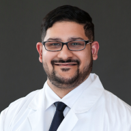
Ziyad Makoshi, MD, MSc, FRCSC, FAANS
Dr. Makoshi completed his neurosurgical residency and general neurosurgery fellowship in Ottawa, Canada, and is a fellow of the Royal College of Physicians and Surgeons of Canada. He completed his American Board of Pediatric Neurosurgery accredited Fellowship at Nationwide Children's Hospital in Columbus, Ohio. He has a Master’s degree in Clinical Neuroscience from University College London, England. He is the author of multiple publications and book chapters on pediatric brain tumors, Chiari malformations, traumatic brain injury, and brain mapping. He is also a member of the Congress of Neurological Surgeons and American Association of Neurological Surgeons. He treats all neurosurgical conditions with particular interest in pediatric brain tumors, epilepsy disorders, hydrocephalus, and cerebral palsy and spasticity. With access to brain mapping software, robotic stereotactic neurosurgery, and the latest technological advances available, Dr. Makoshi and his team provide the highest level of care to children with neurological disorders at El Paso Children’s Hospital.
Sandra Flores, MSN, RN, CPNP-PC, FNP-C, CEN
Ms. Flores is the pediatric neurosurgery nurse practitioner and has over 20 years of nursing experience in multiple care environments with over a decade in emergency and trauma care at a Level 1 trauma
center. She is compassionate and caring and devoted to providing high level patient care to the
underserved population.
Cristobal Perez, PA
Mr. Perez is the pediatric neurosurgery physician assistant with 20 years of experience in the field of
neurosurgery. He brings experience involving pediatrics, neuro-trauma, neuro-intensive care, spine and
neuro-oncology.
Lillian Rodriguez, Neurosciences Coordinator
Tel. (915) 298-5444 ext 80717
Fax. (915) 242-8404
Neurosurgery Team
Dr. John Day, MD
Dr. Luis Vasquez , MD, FACS
Dr. Harold Smith, MD, FAANS
Dr. Jamie Gasco, MD, FAANS, FEBNS
Dr. David Wang, MD, PhD, FAANS
Dr. June Guillet, MD, PhD
Craniofacial Team
Ziyad Makoshi, MD, MSc, FRCSC, FAANS
David Yates, MD, DMD
Kimberly Marquez, RN, BSN Craniofacial Clinical Coordinator
Sandra Flores, CPNP-PC, Pediatric Neurosurgery Nurse Practitioner
Neuroscience Team
Dr. Ziyad Makoshi – Pediatric Neurosurgery
Dr. Rodolfo Fierro-Stevens – Pediatric Neurology
Dr. Violeta Radenovich – Pediatric Ophthalmology
Dr. Sushma Yerram – Epilepsy Neurology
Dr. Alberto Maud – Interventional Neuroradiology
Dr. Gustavo Rodriguez – Interventional Neuroradiology
Dr. Faheem Sheriff – Interventional Neuroradiology
Dr. Vikas Gupta – Interventional Neuroradiology
Dr. Thomas O’Neill – Neuroradiology
Dr. Abraham Lopez – Pediatric Neuropsychology
Epilepsy Team
Dr. Ziyad Makoshi – Pediatric Epilepsy Surgery
Dr. June Gillete – Functional Neurosurgery
Dr. Sushma Yerram – Epilepsy Neurology
Dr. Thomas O’Neill – Neuroradiology
Dr. Abraham Lopez – Pediatric Neuropsychology
Spina Bifida Team
Dr. Ziyad Makoshi – Pediatric Neurosurgery
Dr. Shumyle Alam – Pediatric Urology
Dr. Guillermo Latiff – Pediatric Urology
Dr. Norman Ward – Pediatric Orthopedics
Brachial Plexus Team
Dr. Ziyad Makoshi – Pediatric Neurosurgery
Dr. Shawn Diamond – Plastic Surgery
Contact Us
Lillian E. Rodriguez, Neurosciences Coordinator
lillian.rodriguez@elpasochildrens.org
Office (915) 298-5444 ext 80717
Clinic Phone (915) 242-8402
Clinic Fax (915) 242-8404
Pediatric Neurosurgery – 5400 Alameda Ave., Suite B, El Paso, Texas 79905
For more information please fill out the contact form below
Contact Form:
You are about to leave the El Paso Children’s Hospital Website. El Paso Children’s Hospital is not responsible for the content of the alternate web site. By clicking on the LEARN MORE button below, you acknowledge the previous statement and will be taken to the linked site. If you want to remain at this site, select the CANCEL button.